Turbellarian taxonomic database
Enterostomula evelinae
1948
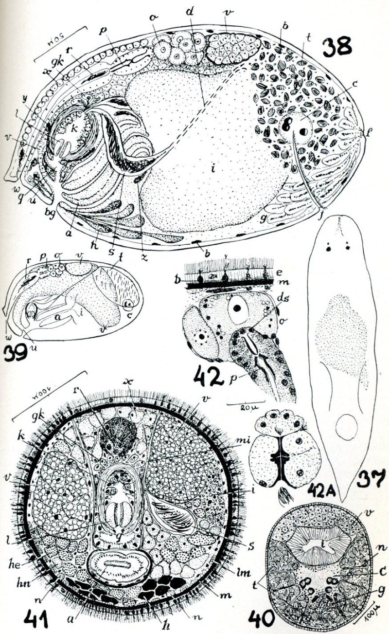
Figs 37-42A
Fig 37: live worm, swimming freely. Fig. 38: reconstruction based on transverse sections. Fig. 39: scheme of organization. Fig 40: transverse section in region of brain. Fig. 41: transverse section in region of male copulatory organ. Fig 42: transverse section in region of ductus spermaticus and two neighboring oocytes Fig. 424A: bursa mouthpiece in a large example. a - pharynx; b - basophilic epidermal secretions; bg - orogenital pore; c - brain; d - efferent duct; ds - internal cells of ductus spermaticus; e - epidermis; f - frontal fossa; g - frontal glands; gk - glands of granular secretions; he - erythrophilic glands; hn - neutrophilic glands; i - gut; j - ciliated groove; k - vesicula granulorum; l - penis; lm - layer of body-wall longitudinal muscles; m - basement membrane; mi - retractora of penis and atrium; n - nerve; o - ovary; p - spermatic duct; q - atrium; r - bursa / in transverse section (Fig 41) vagina externa; s - seminal vesicle; t - testes; u - common ovovitelloduct; v - vitellaria; w - vaginal opening and vagina; x - dorsal glands; y - penial glands; z - pharyngeal glands.
from Marcus Er (1948) Turbellaria do Brasil.
Bol Fac Fil Ci Letr SaoPaulo Zool 13:111-243,22pl
|
