
as Gnathorhynchus hastatus


| Return to Gnathorhynchus hastatus |
D. hastatus
Hochberg, 2004 2004
Fig. 7
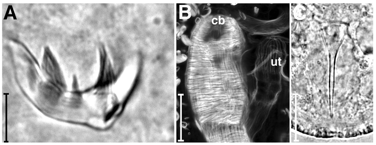
Drepanorhynchides hastatus from Fort Pierce, FL, USA. (A) Light micrograph of proboscis hooks showing three terminal spines or teeth; (B) z-projection of f-actin stained specimen, distal portion of copulatory bulb revealing inner circular muscles of bulb and outer longitudinal muscles; (C) copulatory stylet. cb, copulatory bulb; ut, uterus. Scale bars: A, 10 µm; B, 20µm; C, 25 µm.
|
as Gnathorhynchus hastatus |
|
|
|
|