Turbellarian taxonomic database
Pseudoceros rawlinsonae
2007
Fig 5
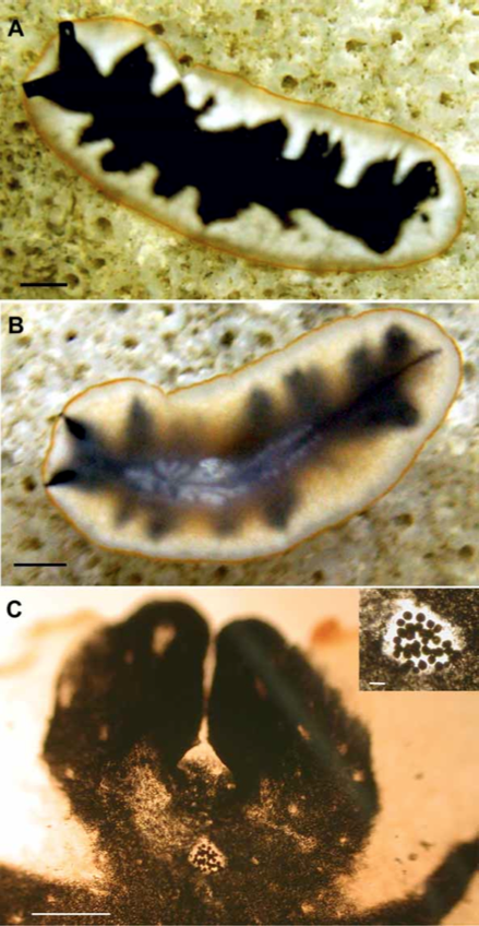
A. Dorsal view of live animal in its natural habitat showing coloration and color pattern. Scale bar 2 mm. B. Ventral view of live animal, showing pharynx and uteri. Scale bar 2 mm. C. Cleared whole mount of the anterior end, showing pseudotentacles and cerebral eye cluster. Scale bar 0.5 mm. Inset: Higher magnification to show detail of cerebral eye cluster. Scale bar 500 μm
from Bolanos DM, Quiroga SY, Litvaitis MK (2007) Five new species of cotylean flatworms (Platyhelminthes: Polycladida) from the wider Caribbean.
Zootaxa 1650:1–23
