Turbellarian taxonomic database
Diascorhynchus falconis
2017
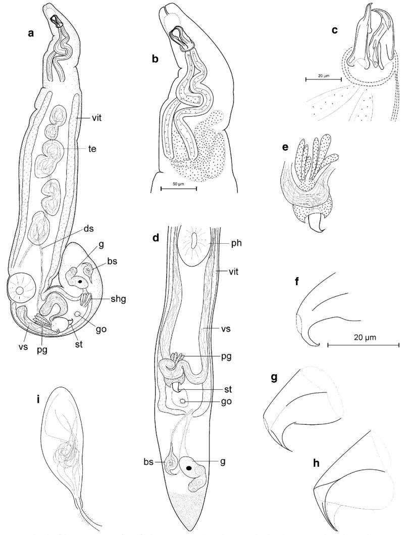
Fig 16
organization from life observation. a) Total view. b) Front end with proboscis, muscular proboscis gland sacs, and gland cells. c) Proboscis. d) Hind end. e) Copulatory organ. f)–h) Stylet. i) Seminal bursa with deferent duct
from Armonies W (2017) Long-term change of meiofaunal species composition in a sandy beach, with description of 7 new species of Platyhelminthes.
Helgoland Marine Research. 71(12). DOI 10.1186/s10152-017-0492-0
