Turbellarian taxonomic database

Bradynectes ensifer
2021
Fig. 1
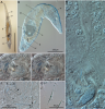
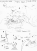
Photomicrographs of Bradynectes ensifer n. sp. using brightfield (A) and DIC (B–G) illumination. A. Living animal, lightly squeezed, dorsal view: brain (b), pharynx (p), gut (g), maturing oocyte (o), seminal vesicle (sv); scale bar = 0.5mm; B. Squeezed specimen, ventral view. Note pharynx pressed open, extension of gut posteriorly beyond maturing oocyte, anterior extent of ovary (ov) lying medially near the posterior end of testis (t), vas deferens (v) and seminal vesicle (sv); C. High-magnification view of male system, from seminal vesicle with mature sperm leading via the intervesicular duct (id) to the prostatic vesicle (pv) with the stylet (st) and surrounded by cement glands (cg); D,E. Ventrally-focused (D) and more dorsally-focused (E) views of the prostatic vesicle and stylet. Note two types of prostatic secretions (ps) and thick muscle (arrow) of the prostatic vesicle that inserts on the anterior part of the stylet base; F. Stylet, showing base opening (bo) and tip opening (to); arrows indicate endpoints for measurement of convex side (cx) and concave side (cv); G. Sperm, released from the seminal vesicle by squeezing;
from Smith JPS, Schärer L (2021) Bradynectes ensifer n. sp. (Platyhelminthes: Macrostomorpha) from North Carolina, USA
Zootaxa 4927 (4): 587–592
| 
|

