Turbellarian taxonomic database
Macrostomum platensis
2012
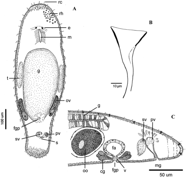
Fig 2
Schematic drawings showing the internal morphology (A) Ventral view of a whole specimen. (B) Stylet from a squash preparation. (C) Reconstruction of the posterior region showing the genital system. cg, cement glands; e, eye; fa, female antrum; fgp, female gonopore; g, gut; m, mouth; mg, male gonopore; oo, oocyte; ov, ovary; pv, prostatic vesicle; rc, rigid cilia; rh, rhabdites; s, stylet; sv, seminal vesicle; t, testes; v, vagina.
from Adami M, Damborenea C, Ronderos JR (2012) A new limnic species of Macrostomum (Platyhelminthes: Macrostomida) from Argentina and its muscle arrangement labeled with phalloidin
Zoologischer Anzeiger 251(3):197-205


