


| Return to Macrostomum platensis |
Macrostomum platensis
2012
Fig 1 D-F
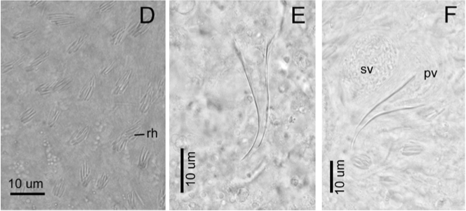
(D) Detailed view of rhabdites at the dorsal surface. (E) Image with higher magnification showing the stylet. (F) Stylet, seminal vesicle and prostatic vesicle. (C)–(F) are interference contrast micrographs.
|
|
|
|