
as Pseudoceros pardalis


| Return to Pseudobiceros pardalis |
Pseudobiceros pardalis
2007
Fig 1 A,B
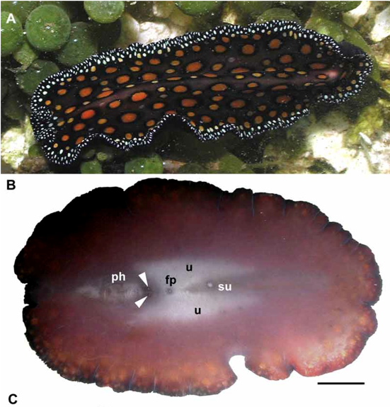
A. Dorsal view of live animal in its natural habitat. Scale bar 5 mm. B. Ventral view of live animal showing pharynx, two male gonopores, female gonopore, uteri and sucker. Scale bar 5 mm.
|
as Pseudoceros pardalis |
|
|