
as Pseudoceros pardalis


| Return to Pseudobiceros pardalis |
Pseudobiceros pardalis
2007
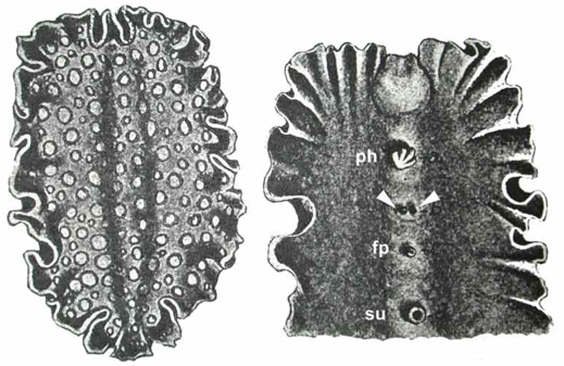
Fig 1 C
Original diagram of Verrill, 1900 of dorsal (left) and ventral view (right); ventral view clearly shows two male gonopores and pharynx, female gonopore and sucker. fp, female gonopore; ph, pharynx; su, sucker; u, uteri; arrowheads, male gonopores.
|
as Pseudoceros pardalis |
|
|